· 3 min read
Advanced Vision for Conventional & Fluorescence Microscopy.
Microscopy is central to biological research and clinical diagnostics — enabling visualisation of structures invisible to the naked eye. Modern vision systems integrate high-resolution cameras, optical sensors, and AI image processing to dramatically improve image quality, sensitivity, and analytical throughput.
Why vision systems matter in microscopy
The quality of microscopy analysis is limited by the quality of image capture and processing. Modern vision systems go far beyond passive image recording — they actively enhance clarity, sensitivity, and analytical speed through AI-driven processing. For fluorescence microscopy in particular, the ability to detect extremely faint emitted light signals determines whether a biological marker can be observed at all.
- 01
Enhanced image clarity
High-resolution cameras capture fine structural details of cells, tissues, and microorganisms — enabling clearer visualisation and more accurate analysis.
- 02
Improved fluorescence sensitivity
Sensitive low-noise detectors capture faint emitted light signals — crucial for observing specific fluorescent molecules or markers at low concentrations.
- 03
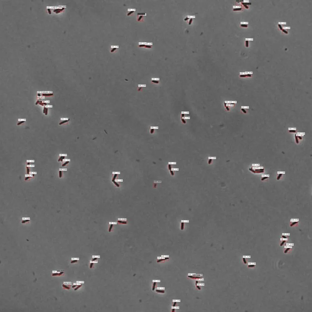
Automated image analysis
AI software automatically identifies, quantifies, and classifies structures — reducing human error, eliminating subjectivity, and dramatically accelerating workflows.
- 04
Real-time live imaging
High-speed cameras enable observation of dynamic biological processes as they happen — essential for cell division, drug response, and fluorescence decay studies.
Applications across fields
Biomedical research — cell behaviour, disease mechanisms, drug interactions, and protein localisation
Clinical diagnostics — detecting pathogens, cancer cells, and biomarkers in patient tissue samples
Pharmaceutical development — screening drug effects at cellular level and quantifying dose-response
Material science — examining microstructures, grain boundaries, and surface defects in advanced materials
Choosing the right vision solution: 8 key factors
Selecting the optimal vision system for microscopy depends on your specific imaging requirements, sample types, and workflow. These are the factors that matter most.
- Type of microscopy <strong>Conventional light microscopy</strong> requires cameras with excellent colour fidelity and high spatial resolution. <strong>Fluorescence microscopy</strong> demands high-sensitivity sensors with low noise to detect weak emitted signals.
- 1
- Type of microscopy Conventional light microscopy requires cameras with excellent colour fidelity and high spatial resolution. Fluorescence microscopy demands high-sensitivity sensors with low noise to detect weak emitted signals.
- Camera sensor technology: CCD vs CMOS CCD sensors offer higher image quality and better sensitivity — ideal for low-light fluorescence imaging. CMOS sensors provide faster frame rates and lower power consumption — preferred for live-cell imaging and video capture. Choose based on whether sensitivity or speed is your primary constraint
- 2
- Camera sensor technology: CCD vs CMOS CCD sensors offer higher image quality and better sensitivity — ideal for low-light fluorescence imaging. CMOS sensors provide faster frame rates and lower power consumption — preferred for live-cell imaging and video capture. Choose based on whether sensitivity or speed is your primary constraint
- Resolution and pixel size Higher resolution cameras with smaller pixel sizes capture finer structural details — but generate larger data files and require more processing power. Balance resolution requirements with your data management and workflow capabilities.
- 3
- Resolution and pixel size Higher resolution cameras with smaller pixel sizes capture finer structural details — but generate larger data files and require more processing power. Balance resolution requirements with your data management and workflow capabilities.
- Sensitivity and dynamic range Sensitivity is critical in fluorescence microscopy where emitted signals are extremely faint. High quantum efficiency and low read noise improve signal detection. Dynamic range allows distinguishing very bright and very dim features within the same image. Most important factor for fluorescence applications
- 4
- Sensitivity and dynamic range Sensitivity is critical in fluorescence microscopy where emitted signals are extremely faint. High quantum efficiency and low read noise improve signal detection. Dynamic range allows distinguishing very bright and very dim features within the same image. Most important factor for fluorescence applications
- Frame rate High frame rates are essential for live imaging — capturing fast biological processes without motion blur. Particularly relevant for time-lapse microscopy, calcium imaging, and tracking rapidly moving cells.
- 5
- Frame rate High frame rates are essential for live imaging — capturing fast biological processes without motion blur. Particularly relevant for time-lapse microscopy, calcium imaging, and tracking rapidly moving cells.
- Image processing and software integration Advanced vision solutions include software for real-time image enhancement, noise reduction, and automated analysis — cell counting, fluorescence quantification, and morphological classification. Compatibility with existing microscopy setups and data management systems is essential.
- 6
- Image processing and software integration Advanced vision solutions include software for real-time image enhancement, noise reduction, and automated analysis — cell counting, fluorescence quantification, and morphological classification. Compatibility with existing microscopy setups and data management systems is essential.
- Environmental conditions Some applications require temperature-controlled or vibration-isolated setups. The vision system must be robust and adaptable to these conditions for consistent performance across imaging sessions.
- 7
- Environmental conditions Some applications require temperature-controlled or vibration-isolated setups. The vision system must be robust and adaptable to these conditions for consistent performance across imaging sessions.
- Budget and scalability Consider initial investment alongside ongoing costs — maintenance, software licences, and future upgrades. Choose solutions that can evolve with your research needs rather than requiring full replacement as requirements change.
- 8
- Budget and scalability Consider initial investment alongside ongoing costs — maintenance, software licences, and future upgrades. Choose solutions that can evolve with your research needs rather than requiring full replacement as requirements change.
Key benefits of advanced microscopy vision systems
Higher image quality and analytical accuracy across conventional and fluorescence modalities
Automated analysis reducing human error and dramatically accelerating research throughput
Improved sensitivity for low-light fluorescence detection — seeing markers that weaker systems miss
Real-time live imaging capability for dynamic biological process observation
- 01
Key capabilities
Modalities Light + Fluorescence Analysis Automated Live imaging Real-time Sensor options CCD / CMOS Integration MES / LIMS
- 02
Application areas
Biomedical research Clinical diagnostics Pharma screening Material science QC / Inspection
- 03
Related guides
Medical device testing Pharma glass inspection Custom AI services
Related reading
More guides across labs, pharma, and life sciences.
Guide · Medical Automated Testing of Medical Devices Read more
Guide · Pharma Computer Vision for Glass & Vial Inspection Read more
Guide · Pharma
Vision Inspection for Pharmaceutical Packaging
Read more